Science / Grade 6
Kidneys: filter, return and remove
Learning goal: Separate first filtering from reabsorption, follow blood and urine routes, and repair a museum model with clear arrow labels.
Before you start: Follow a labeled arrow and distinguish blood vessels from the digestive tube. Review the oxygen and digestion routes when needed. Use only the supplied models, not personal health observations.
Read or print this lesson What to practiceThe video could not load. The transcript is still available below.
Next: your worksheet
Kidneys: Filter Is Not Discard
Checking sign-in...
Read the transcript
Saved reading place
Video transcript and practice. Reading or printing does not count as playback time or an assessed grade.
1. A filter is only the beginning
Video: 0:00
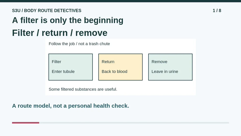
A museum robot has a terrible sorting plan: anything that passes through a filter must go in the trash. Even its lunch ticket. Let us improve that plan using a kidney model. Kidneys help remove wastes and extra water from blood, forming urine. But filtering is not the entire job. Much of the filtered water and needed substances return to blood. We will follow the different arrows, then repair the robot's labels. Use the supplied paper models only. No body measurements, samples, drinking experiment, or personal health information are needed.
2. Blood and urine have different routes
Video: 0:44
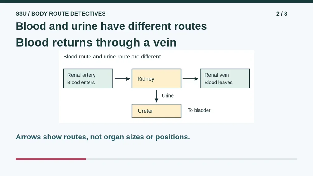
Start with the blood route. Blood enters a kidney through a renal artery, travels through smaller vessels, and leaves through a renal vein. Renal simply means related to the kidney. Now find the separate urine route. Urine formed in the kidney travels toward the bladder through a tube called a ureter. The vein and ureter do not carry the same liquid. Do not make every arrow mean urine, and do not send blood into the bladder. Our boxes show connections, not the actual shapes, sizes, or locations of the organs.
3. What crosses the first filter?
Video: 1:22
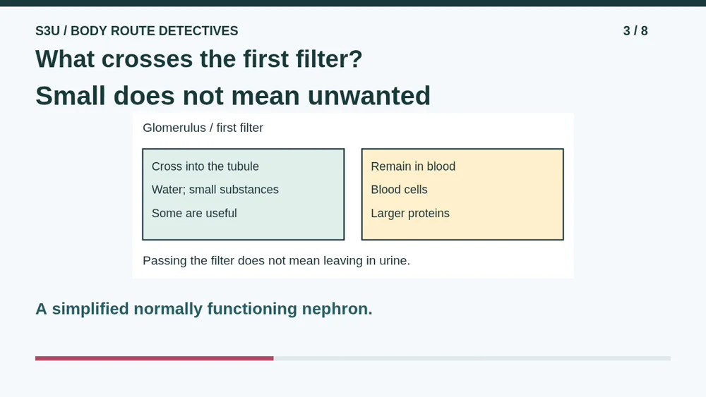
Look inside one tiny working unit called a nephron. Its first filter is a cluster of small blood vessels called the glomerulus. In this simplified normally functioning model, water and small dissolved substances pass from blood into the tubule, a tiny tube. Blood cells and larger proteins remain in the blood vessels. Notice an important surprise: some filtered substances are useful. Passing the first filter does not mean a substance is waste or that it will leave the body. The robot needs a second step before it makes its final decision.
4. Return useful substances to blood
Video: 2:04

As filtered fluid travels through the tubule, much water and needed substances return to nearby blood. That return is called reabsorption. Follow the direction carefully: from fluid in the tubule back into blood, not from the bladder back to the kidney. The tubule also helps move additional wastes from blood into its fluid. What remains becomes urine. Our two arrows show different directions, not identical actions. Real kidneys regulate these movements; a kitchen sieve alone cannot explain them. The model does not say that every small substance is kept or every filtered substance is discarded.
5. The bladder stores; it does not filter
Video: 2:49
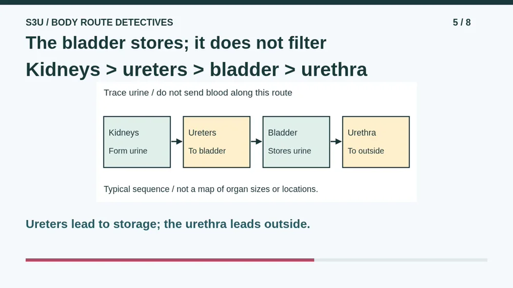
Now trace urine after it has been formed. It travels from the kidneys through the ureters to the bladder. The bladder stores urine until it leaves through the urethra. Ureter and urethra sound similar, so use their destinations to tell them apart. The ureters lead from kidneys to bladder; the urethra leads from bladder to outside. Storage is not blood filtering. Also keep this route separate from the digestive tube: stool leaves by a different route. These diagrams describe a typical learning model, not the exact anatomy or health of every person.
6. Pause: repair the robot labels
Video: 3:31

Pause for two fresh labels at the museum. Label A says, a useful dissolved substance crossed the first kidney filter, so it must leave in urine. Label B says, urine travels from a kidney through the urethra to the bladder, where blood is filtered. Repair both claims. For A, explain what could happen after the first filter. For B, name the tube to the bladder and the bladder's job. Then name the route from the bladder to outside. You may point, speak, or write. Continue when you have reasons, not just replacement words.
7. Check the direction and the job
Video: 4:11
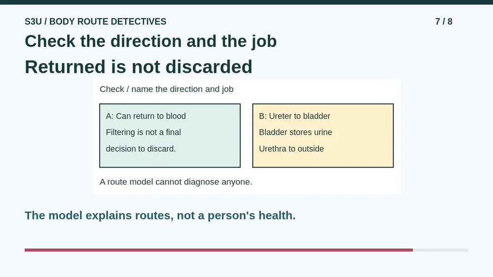
For A, a needed substance can return from the tubule fluid to blood. Crossing the first filter does not guarantee that it will leave in urine. For B, a ureter carries urine from a kidney to the bladder. The bladder stores urine; kidneys filter blood. The urethra carries urine from the bladder to outside. Our robot must read both the arrow direction and its label. One arrow is not interchangeable with another. These paper routes cannot diagnose a person or supply an exact amount of urine. No personal observations are necessary to explain the repairs.
8. Tiny units, connected jobs
Video: 4:55
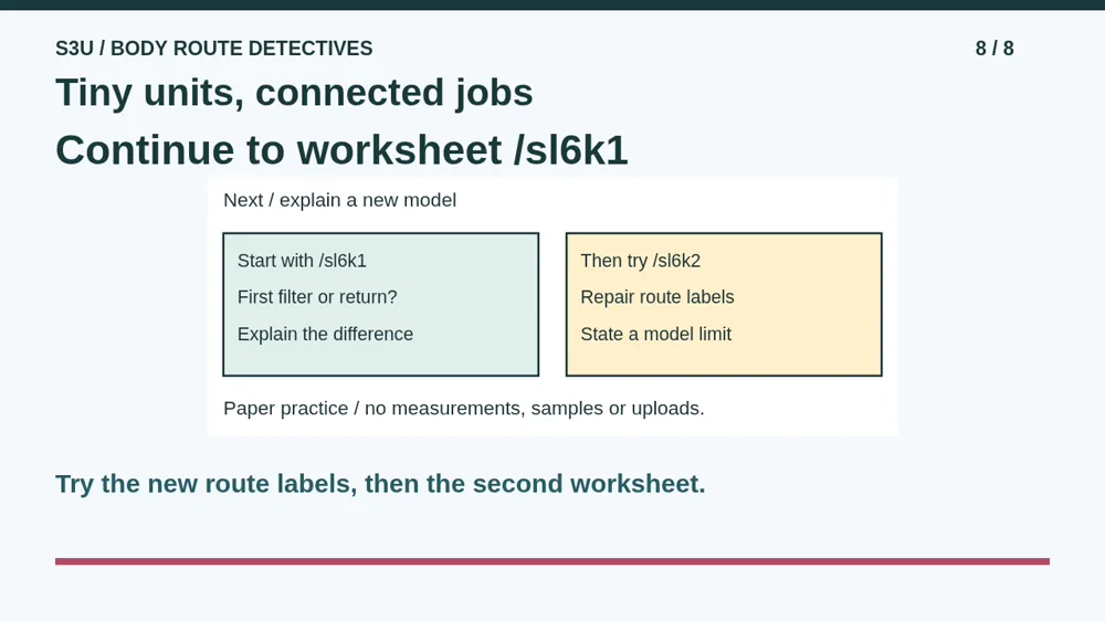
Fun fact: a kidney contains about a million tiny filtering units called nephrons. That is an approximate count, not a promise that every kidney is identical. Each unit connects filtering with further work along a tubule. The museum robot has plenty to learn from that teamwork. Continue to the first worksheet and distinguish what stays in blood, what crosses, and what can return. Then use the second worksheet to repair different route labels. Explain at least one limit of the drawings. You can complete this practice on paper without an account or an upload.
Show your understanding
You can point, explain aloud, draw or write.
- Distinguish first filtering into a nephron tubule from returning needed substances to blood.
- Trace urine through ureters, bladder and urethra while keeping blood and stool routes separate and explaining a drawing limit.
Try it yourself
Pause at the two robot labels. Explain why crossing the first filter does not guarantee leaving in urine, then repair the tube and storage claims.
Continue to both worksheets with their new arrow labels. Use the supplied models only; no personal health information or body measurements are needed.
Next: your worksheet
Kidneys: Filter Is Not Discardhttps://s3u.com/sl6k1
Lesson: https://s3u.com/lessons/filter-and-return